Poster Presentation Australian Diabetes Society and the Australian Diabetes Educators Association Annual Scientific Meeting 2014
Phenotypic Changes Of Tissue Macrophage Populations In Mouse Models of Diabetes and Obesity (#251)
Macrophage infiltration to tissue is a characteristic feature of many diabetic complications. Diabetes and obesity are both characterised by an increase in systemic inflammation. Whether this increase in inflammation can affect tissue macrophage populations is unknown and was investigated in murine models of diabetes and obesity in this study.
At 5 weeks of age C57BL/6 mice were made diabetic using streptozotocin (3x55mg/kg). Similarly aged mice fed either a HFD (45%kCal fat) or chow acted as control. Animals (n=4-8/grp) were terminated after 5, 10 and 20 weeks and kidneys were harvested for qRT-PCR analysis of macrophage subpopulations (tissue macrophage F4/80, M1:CD11c and M2:CD163) reparative to F4/80/CD11c positive (M1 polarised ) and F4/80/CD163 positive (M2 polarised).Fibrosis markers (Collagen I (Col 1), Fibronectin (Fn) and Collagen IV (Col IV)) were also investigated. Urine and blood were also collected.
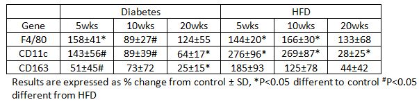
Diabetes caused a transient induction of F4/80 gene expression and decreased expression of CD11c and CD163, reaching significance at 20 weeks. Similarly HFD increased expression of F4/80 and CD11c which persisted until 10 weeks and for CD11c was of greater magnitude (HFD: 2.7 fold vs DM: 0.9 fold at 10 weeks). In contrast CD163 levels in HFD were unaltered. These changes preceded induction of fibrosis markers and alterations in kidney function (not shown).
Diabetes and high fat diet had no effect on kidney macrophage numbers as measured by F4/80 but the expression of both M1 and M2 markers decreased albeit at different rates. How these changes in macrophage phenotype relate to complications development remains to be further studied